Parasitic Protozoa of Farm Animals and Pets by Monica Florin-Christensen & Leonhard Schnittger
Author:Monica Florin-Christensen & Leonhard Schnittger
Language: eng
Format: epub
Publisher: Springer International Publishing, Cham
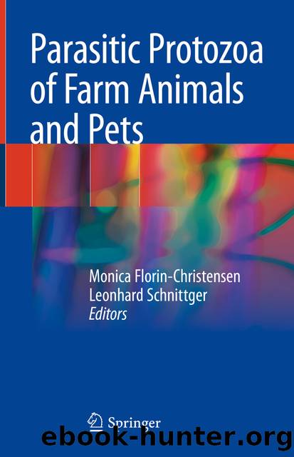
As shown in Fig. 9.2, Babesia merozoites share with other eukaryotic cells the presence of membranous organelles, such as nucleus, endoplasmic reticulum (ER), Golgi apparatus, and mitochondria. In addition, as other Alveolata members, they are characterized by the presence of a cellular wrapping—the pellicle—which is composed of the cell membrane and two inner layers: a primary layer of vesicles or alveoli and a secondary layer of microtubules, involved in parasite motility and host cell invasion (Lew et al. 2002). As in other Apicomplexans, an assembly of invasion-specialized organelles—the apical complex—is present at the anterior apex. It is composed of a polar ring, micronemes, and rhoptries. In contrast, another typical apical structure—the conoid—is absent in piroplasmids (Blackman and Bannister 2001; Klinger et al. 2013). Spherical bodies are unique secretory organelles of Babesia spp. distributed in the parasite cytoplasm. They are considered to be homologous to the dense granules, present in other apicomplexans, and to participate in host-pathogen interactions during the intraerythrocytic stage. An apicoplast, a plastid with no photosynthetic activity, thought to have been acquired by secondary endosymbiosis from algae, also occurs in these cells. The apicoplast takes care of some metabolic activities, such as fatty acid and isoprenoid biosynthesis (Caballero et al. 2012). Due to its essential functions for parasite survival, this special plastid has gained attention as an attractive target for the development of parasiticidal drugs (Brayton et al. 2007; Huang et al. 2015).
Fig. 9.2Morphology of a Babesia merozoite. Organelles shared with other eukaryotes, other Alveolata and other Apicomplexa are shown. A conoid is not present in Babesia apical complex. Spherical bodies of this parasite are homologous to apicomplexan-typical dense granules. They do not co-localize with the apical complex but are functionally associated
Download
This site does not store any files on its server. We only index and link to content provided by other sites. Please contact the content providers to delete copyright contents if any and email us, we'll remove relevant links or contents immediately.
Periodization Training for Sports by Tudor Bompa(8368)
Why We Sleep: Unlocking the Power of Sleep and Dreams by Matthew Walker(6811)
Paper Towns by Green John(5269)
The Immortal Life of Henrietta Lacks by Rebecca Skloot(4667)
The Sports Rules Book by Human Kinetics(4478)
Dynamic Alignment Through Imagery by Eric Franklin(4305)
ACSM's Complete Guide to Fitness & Health by ACSM(4110)
Kaplan MCAT Organic Chemistry Review: Created for MCAT 2015 (Kaplan Test Prep) by Kaplan(4075)
Livewired by David Eagleman(3847)
Introduction to Kinesiology by Shirl J. Hoffman(3829)
The Death of the Heart by Elizabeth Bowen(3697)
The River of Consciousness by Oliver Sacks(3663)
Alchemy and Alchemists by C. J. S. Thompson(3575)
Bad Pharma by Ben Goldacre(3490)
Descartes' Error by Antonio Damasio(3356)
The Emperor of All Maladies: A Biography of Cancer by Siddhartha Mukherjee(3232)
The Gene: An Intimate History by Siddhartha Mukherjee(3155)
The Fate of Rome: Climate, Disease, and the End of an Empire (The Princeton History of the Ancient World) by Kyle Harper(3109)
Kaplan MCAT Behavioral Sciences Review: Created for MCAT 2015 (Kaplan Test Prep) by Kaplan(3046)
