Grabb and Smith's Plastic Surgery (GRABB'S PLASTIC SURGERY) by Thorne Charles H
Author:Thorne, Charles H. [Thorne, Charles H.]
Language: eng
Format: epub
Publisher: Lippincott Williams & Wilkins
Published: 2013-09-26T00:00:00+00:00
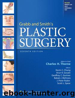
FIGURE 47.2. The anatomic layers of the face. Although the quality of the layers differs in various areas of the face, the arrangement of layers is identical. The facial nerve (cranial nerve [CN] VII) branches innervate their respective muscles via their deep surfaces.
Facial Nerve
If the surgeon remembers that the facial nerve branches innervate the respective facial muscles via their deep surfaces, the safe planes of dissection become obvious. Dissection in the subcutaneous plane, superficial to the SMAS–muscle layer, is safely performed anywhere in the face, whether it is the temporal region, cheek, or neck. Dissection deep to the SMAS, superficial to the facial nerve branches, requires care.
There are three to five frontal (or temporal) branches of the facial nerve that cross the zygomatic arch and innervate the frontalis muscle, orbicularis oculi, and corrugator muscles via their deep surfaces.3 Because the layers of anatomy, although present, are compressed over the arch, these branches are vulnerable to injury in this region. Dissection in this region can either be performed superficial to the nerve branches in the subcutaneous plane, or deep to the branches on the surface of the temporalis muscle fascia (deep temporal fascia).4
The zygomatic branches innervate the orbicularis oculi and zygomaticus muscles. One must remember that although the facial nerve branches travel deep to the SMAS layer, at some point these branches turn superficially to innervate the overlying muscles. Any dissection in the sub-SMAS plane in the cheek, whether as part of a composite rhytidectomy or standard dissection of the SMAS as a separate layer, necessitates a change of surgical planes at the zygomaticus major muscle to avoid transection of the branch to this muscle. The dissection plane changes from sub-SMAS to subcutaneous by passing over the superficial surface of the zygomaticus major and thereby preserving its innervation.
The buccal branches lie on the masseter muscle and are easily visualized through the parotid–masseteric fascia. Some buccal branches merge with branches of zygomatic origin to innervate the procerus muscle and provide additional innervation of the corrugator muscle. Consequently, the corrugator muscle receives innervation from the frontal, zygomatic, and buccal branches.
Earlier publications indicated that the marginal mandibular branches were located above the inferior border of the mandible in many cases. More recent studies demonstrate that, in fact, these branches are always located caudal to the inferior border of the mandible. The cervical branches innervate the platysma muscle.
Anatomic studies indicate that there are fewer crossover communications between the frontal branches and marginal mandibular branches, which helps to explain why injuries to these nerves are less likely to recover function in their respective muscles than injuries to the zygomatic or buccal branches.
Retaining Ligaments
In at least two areas of the face the anatomic layers are condensed and less mobile with respect to each other. These “ligaments” are areas where the skin and underlying tissues are relatively fixed to the bone.5 The zygomatic ligament (previously known as the McGregor patch) is located in the cheek, anterior and superior to the parotid gland, and posteroinferior to the malar eminence.
Download
This site does not store any files on its server. We only index and link to content provided by other sites. Please contact the content providers to delete copyright contents if any and email us, we'll remove relevant links or contents immediately.
| Anesthesiology | Colon & Rectal |
| General Surgery | Laparoscopic & Robotic |
| Neurosurgery | Ophthalmology |
| Oral & Maxillofacial | Orthopedics |
| Otolaryngology | Plastic |
| Thoracic & Vascular | Transplants |
| Trauma |
Periodization Training for Sports by Tudor Bompa(8274)
Why We Sleep: Unlocking the Power of Sleep and Dreams by Matthew Walker(6726)
Paper Towns by Green John(5191)
The Immortal Life of Henrietta Lacks by Rebecca Skloot(4589)
The Sports Rules Book by Human Kinetics(4388)
Dynamic Alignment Through Imagery by Eric Franklin(4217)
ACSM's Complete Guide to Fitness & Health by ACSM(4060)
Kaplan MCAT Organic Chemistry Review: Created for MCAT 2015 (Kaplan Test Prep) by Kaplan(4014)
Introduction to Kinesiology by Shirl J. Hoffman(3776)
Livewired by David Eagleman(3775)
The Death of the Heart by Elizabeth Bowen(3622)
The River of Consciousness by Oliver Sacks(3605)
Alchemy and Alchemists by C. J. S. Thompson(3522)
Bad Pharma by Ben Goldacre(3428)
Descartes' Error by Antonio Damasio(3279)
The Emperor of All Maladies: A Biography of Cancer by Siddhartha Mukherjee(3163)
The Gene: An Intimate History by Siddhartha Mukherjee(3099)
The Fate of Rome: Climate, Disease, and the End of an Empire (The Princeton History of the Ancient World) by Kyle Harper(3067)
Kaplan MCAT Behavioral Sciences Review: Created for MCAT 2015 (Kaplan Test Prep) by Kaplan(2987)
