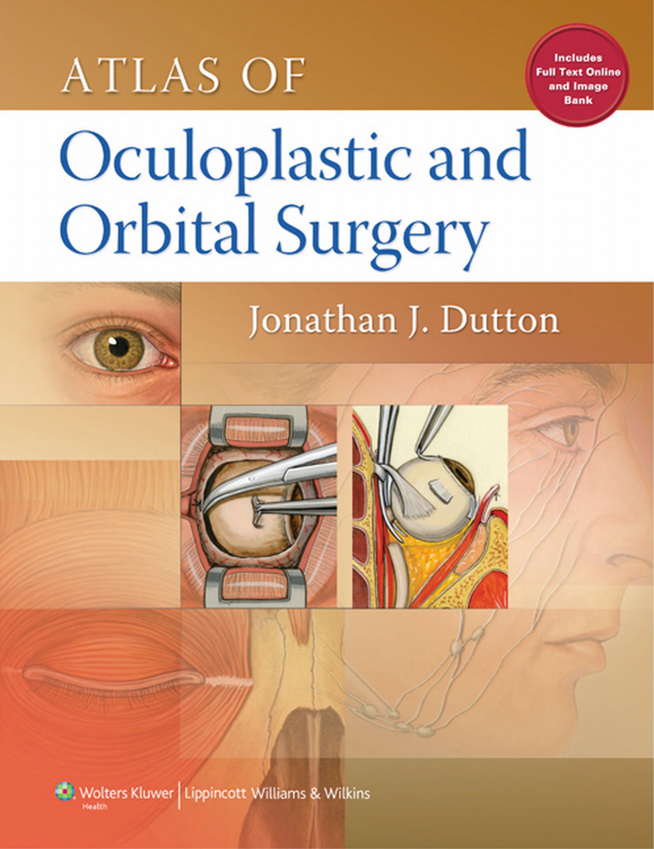
Atlas of Oculoplastic and Orbital Surgery by Jonathan Dutton

Author:Jonathan Dutton
Language: eng
Format: epub, pdf
ISBN: 9781469817538
Publisher: Lippincott Williams & Wilkins
Upper Eyelid Reconstruction
Full-thickness defects of the upper eyelid may result from mechanical or thermal trauma, from the surgical excision of eyelid tumors, or from congenital colobomas. In the reconstruction of such defects, it is essential not only to reestablish the anatomic integrity of the lid but also to restore its physiologic function. The surgeon must pay special attention to the individual layers of the eyelid to ensure appropriate mobility and protection of the globe.
Small full-thickness marginal defects of 25% to 30% may be closed by direct layered closure, depending on the laxity of the eyelid. In older individuals, when sufficient eyelid laxity permits, defects of 40% or more may be repaired by this technique. The functional and cosmetic results are superior to any other procedure, and when it is performed properly, direct layered closure leaves an intact lid margin with a full lash line.
In more complicated reconstructive procedures, the anterior lamella of the upper lid must be covered with thin skin, which is loose enough to allow complete eyelid closure, yet thin and flexible enough to fold easily when the lid is opened. A circumferential muscle layer is necessary for closure to prevent lagophthalmos and corneal exposure. Vertical retraction, either with the levator muscle or a suitable substitute, is necessary to elevate the lid above the visual axis. Internal support by replacement of the tarsus or other firm tissue provides marginal stability and intimate corneal contact in all positions of gaze. Reconstruction of the canthal ligaments is less important here than in the lower eyelid because the effects of gravity enhance eyelid position rather than oppose it. A mucous membrane lining on the posterior eyelid surface is critical to prevent corneal abrasion. Meticulous detail is paid to reconstruction of the eyelid margin to exclude keratinized epithelium and prevent notching and trichiasis.
Many procedures are available for the partial or complete reconstruction of the upper eyelid. The choice depends upon numerous factors, and frequently a combination of techniques is necessary for adequate repair. In traumatic injuries, especially following thermal or chemical burns, tissue vascularity may be compromised. In such situations, free grafts may not take as readily; therefore, the use of a vascularized flap may be more appropriate. The same is true for heavily irradiated tissues. The development of local flaps for eyelid reconstruction requires some degree of tissue laxity, which may not readily be available in younger individuals or in those with cicatrizing skin diseases.
No hard and fast rules for the reconstruction of specific defects can be given. The surgical approach is dictated by the size and location of the defect; involvement of deep eyelid structures, such as the levator aponeurosis or canthal ligaments; the availability of adjacent or distant tissue for repair; and the skills of the surgeon. Careful evaluation of the defect and essential functional components of the eyelid must precede any attempt at repair. Basic techniques are illustrated below, but the appropriate application of these procedures, in combination when necessary, will determine the final functional and cosmetic result.
When
Download
Atlas of Oculoplastic and Orbital Surgery by Jonathan Dutton.pdf
This site does not store any files on its server. We only index and link to content provided by other sites. Please contact the content providers to delete copyright contents if any and email us, we'll remove relevant links or contents immediately.
| Anesthesiology | Colon & Rectal |
| General Surgery | Laparoscopic & Robotic |
| Neurosurgery | Ophthalmology |
| Oral & Maxillofacial | Orthopedics |
| Otolaryngology | Plastic |
| Thoracic & Vascular | Transplants |
| Trauma |
When Breath Becomes Air by Paul Kalanithi(7256)
Why We Sleep: Unlocking the Power of Sleep and Dreams by Matthew Walker(5637)
Paper Towns by Green John(4165)
The Immortal Life of Henrietta Lacks by Rebecca Skloot(3821)
The Sports Rules Book by Human Kinetics(3582)
Dynamic Alignment Through Imagery by Eric Franklin(3483)
ACSM's Complete Guide to Fitness & Health by ACSM(3462)
Kaplan MCAT Organic Chemistry Review: Created for MCAT 2015 (Kaplan Test Prep) by Kaplan(3419)
Introduction to Kinesiology by Shirl J. Hoffman(3297)
Livewired by David Eagleman(3117)
The River of Consciousness by Oliver Sacks(2989)
Alchemy and Alchemists by C. J. S. Thompson(2909)
The Death of the Heart by Elizabeth Bowen(2897)
Descartes' Error by Antonio Damasio(2728)
Bad Pharma by Ben Goldacre(2724)
The Gene: An Intimate History by Siddhartha Mukherjee(2489)
Kaplan MCAT Behavioral Sciences Review: Created for MCAT 2015 (Kaplan Test Prep) by Kaplan(2486)
The Fate of Rome: Climate, Disease, and the End of an Empire (The Princeton History of the Ancient World) by Kyle Harper(2431)
The Emperor of All Maladies: A Biography of Cancer by Siddhartha Mukherjee(2427)
