SAFE MICROSCOPIC TECHNIQUES FOR AMATEURS Slide Mounting by Walter Dioni
Author:Walter Dioni [Dioni, Walter]
Language: eng
Format: epub
Published: 2014-06-01T07:00:00+00:00
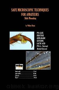
CHAPTER 3A: Formulae Derived From Fructose FG—FructoseGlycerol Medium Brun’s medium was formulated to use glucose as the “sugar” ingredient. I have modified the formula to replace the glucose with fructose (in my case, Karo syrup).
Fructose at a 76% concentration 24 ml
Glycerol 3 ml
Antiseptic (I use Listerine) 3 ml
2
1 The first picture [1] shows the
surface of a fly wing, and the
second [2] the border of the
same. The picture [3] is from
the epithelium of the underside
of a leaf, showing one stomata
and the nuclei of two marginal
cells. The fourth (opposite
page) is the compound image
of the 5th leg from a female of
a species of Diaptomus, a
genus of copepod
(crustaceans). The sixth is the
ovisac (a sac in which the 3 female carries the eggs). All
were taken of slides mounted in
FG. The wings were stored in alcohol 70%. The epithelia was fixed for
5 24 hrs in AFA (a fi xative composed of Alcoh o l, Formalin, and Acetic acid) and the picture taken with the 100x object i v e (1000x), The copepods were fixed more than a month ago in lactocupric, a
fixative that I will discuss in a future article, and this specimen was washed in water for ten minutes and directly transferred to the mounting media. As we will see in other images, this is responsible for the collapse of the eggs. Some intermediate steps through glycerinated water can prevent this.
6
A modification that is also working for me is the addition of lactic acid as a clearing agent
FGL - Modified Brun’s medium Fructose (76% concentration) 21 ml Glycerol 3 ml Lactic acid 3 ml Antiseptic 3 ml
These two media, give good results with the test objects but take longer to dry. I estimate the drops so they don’t exceed the coverslip, and twenty four hours later I seal, with the same care as with a glycerin mountant. If not well sealed the coverslip continues to be "slippery" for more than a week.
The following images show examples. The first two [wing] & [epit] are similar to the FG pictures. The Diaptomus female in the third picture [ovisac] has adhered the ovisac, and also two spermatophores (sperm packets). Comments as in the FG case. The last three pictures are from a mosquito larva, fixed in alcohol 70% and first cleared in lactoglycerol, before being mounted in FGL. The head shows clearly the interior structure with the brain lobules, the nerves connecting to the eyes, several muscles and other sensitive organs. The ventral side of the head shows part of the buccal armature. The air tube is part of the caudal complex I depicted and labelled in the first article; you may wish to reread as a reminder of the anatomical features.
FGL, wing
FGL, epit FGL, ovisac
FGL, larva head FGL, telson setae
FGL, ventral head
Please refer to text for imageFGL, airtube
Download
This site does not store any files on its server. We only index and link to content provided by other sites. Please contact the content providers to delete copyright contents if any and email us, we'll remove relevant links or contents immediately.
| Electron Microscopes & Microscopy | Experiments & Projects |
| Measurement | Microscopes & Microsocopy |
| Scientific Instruments | Telescopes |
| Time | Methodology & Statistics |
Hands-On Genetic Algorithms with Python by Eyal Wirsansky (2020) by Unknown(4140)
Thing Explainer by Randall Munroe(3996)
The Elements by Theodore Gray(3136)
The Meaning of it All by Richard Feynman(2396)
Make by Mike Westerfield(2350)
Every Tool's a Hammer by Adam Savage(1998)
Science Experiments You Can Eat by Vicki Cobb(1918)
The Perfectionists by Sara Shepard(1859)
Martin Gardner's Science Magic by Martin Gardner(1772)
Raspberry Pi Electronics Projects for the Evil Genius (Tab) by Norris Donald & Norris Donald(1729)
Handbook of Modern Sensors by Jacob Fraden(1702)
Synchrotron Light Sources and Free-Electron Lasers by Eberhard J. Jaeschke Shaukat Khan Jochen R. Schneider & Jerome B. Hastings(1677)
Elephants on Acid by Boese Alex(1631)
Elephants on Acid: And Other Bizarre Experiments by Alex Boese(1613)
The Perfectionists by Simon Winchester(1603)
Tesla by Carlson W. Bernard(1557)
The Science of Food by Marty Jopson(1502)
The Meaning Of It All by Richard P. Feynman(1486)
125 Physics Projects for the Evil Genius by Silver Jerry(1477)
