Make Life Visible by Unknown
Author:Unknown
Language: eng
Format: epub
ISBN: 9789811379086
Publisher: Springer Singapore
15.3 Progress in Vascular Imaging of Small Animals with Lab Systems
Preclinical microCT systems are commercially available that enable CT angiography in animals up to the size of rabbits. However, effective pixel size for single projection images on the CMOS flat-panel detectors with a field of view of several cm3 is typically 100–200 μm. With such systems 2D angiography can at best reveal small arteries. Higher resolution acquisitions are only possible for ex vivo 3D acquisitions due to the prolonged scanning times (>5 min). Nonetheless, microfocus X-ray imaging systems are available that are suitable for real time imaging of the microvessels in vivo, but the challenge has been achieving fast imaging with sufficient absorption contrast to visualize vessels in organs that move, the heart and lungs. One such system that we have utilized for investigations of the hindlimb vasculature in peripheral arterial disease associated with diabetes is the Hitex system described in our recent study (Sonobe et al. 2015).
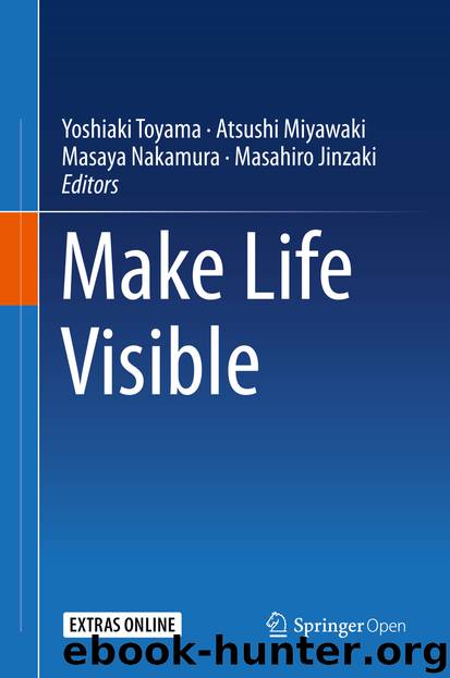
Utilising an optimised microfocus X-ray system (MFX-80HK, Hitex Ltd., Osaka, Japan) incorporating an off the shelf high speed camera with ultrasensitivity (FASTCAM Mini AX200, Photron, Japan) we have greatly improved the possibilities for real time cine-angiography in rats and mice (Fig. 15.6). The current capability of this system suggests that microvessel function studies approach the real resolution of SR microangiography in the hindlimb, brain and renal vessel beds (with frame averaging). However, limited photon counts at the detector result in blur and unsatisfactory vessel edge detection in single projection images of the coronary and pulmonary arteries below 100 μm. Further optimization of the image intensifier and X-ray source might lead to the development of a system that permits assessment of coronary endothelial function in vivo during closed-chest conditions in the laboratory.
Fig. 15.6(a) Microfocus X-ray video system (Hitex, Osaka, Japan) optimized for laboratory based imaging (iodine contrast agent) and examples of image quality achieved while imaging anaesthetised (b) rats (hindlimb, kidney, pulmonary and coronary arteries) and (c) mice (hindlimb), acquired at 250 frames/s, 60 kV and 80 μA. Black arrows indicate 50 μm arterioles in the hindlimb and renal cortex
Download
This site does not store any files on its server. We only index and link to content provided by other sites. Please contact the content providers to delete copyright contents if any and email us, we'll remove relevant links or contents immediately.
4 - Harry Potter and the Goblet of Fire by J.K. Rowling(2874)
2010-The City & the City by China Miéville(2046)
Wall and Piece by Banksy(1901)
Thank You for Being Late by Thomas L. Friedman(1826)
The Old Farmer's Almanac 2020 by Old Farmer’s Almanac(1823)
The Mirror and the Light by Hilary Mantel(1746)
Journey to the Abyss by Harry Kessler(1692)
The Andy Warhol Diaries by Andy Warhol(1671)
Tolkien, J. R. R. - The Fellowship of the Ring by Tolkien J. R. R(1611)
Life on Earth by David Attenborough(1583)
Art Nouveau by Carol Belanger Grafton(1485)
The Museum Of Innocence by Orhan Pamuk(1476)
Harry Potter - A History of Magic by British Library(1468)
The Business of Being an Artist by Daniel Grant(1434)
The Future Is Japanese by(1410)
Frida by Hayden Herrera(1377)
Impeach by Neal Katyal(1344)
A History of Japanese Art by Noritake Tsuda(1283)
Insight Guides Japan (Travel Guide eBook) by Insight Guides(1256)
